
Mesenchymal Stem Cells in Neurological Disease: Paracrine Mechanisms and Clinical Application
Dr. Uladzislau Tsvirko
12 March 2025
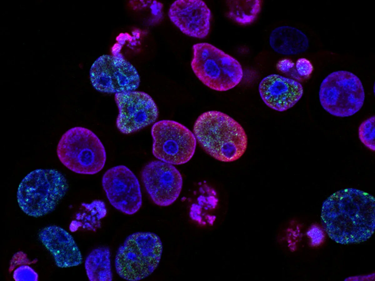
MSCs exert their therapeutic effect not by replacing damaged tissue, but through paracrine signalling — secreting anti-inflammatory cytokines, trophic factors, and extracellular vesicles that modulate the disease microenvironment.
Beyond Cell Replacement
The early hypothesis that mesenchymal stem cells would differentiate into replacement neurons has largely been superseded by a more nuanced understanding. MSCs exert their primary therapeutic effect through paracrine signalling — the secretion of bioactive molecules that modulate the local microenvironment surrounding diseased tissue. This mechanism, rather than direct cell replacement, explains why MSC therapy demonstrates efficacy across biologically distinct conditions: from motor neuron disease to autoimmune demyelination to progressive cerebellar degeneration.
Molecular Secretome
MSCs produce a complex secretome comprising anti-inflammatory cytokines (IL-10, TGF-β), neurotrophic factors (BDNF, GDNF, VEGF), and extracellular vesicles. This molecular output collectively suppresses pathological immune activation, reduces oxidative stress, promotes angiogenesis, supports neuronal and glial cell survival, and modulates the local microenvironment to favour endogenous repair. The breadth of this secretome explains the multi-target nature of MSC therapy — a characteristic that single-molecule pharmaceutical agents cannot replicate.
Sourcing and Characterisation
At BioCells Medical, MSCs are isolated from the patient’s own bone marrow (autologous, approximately 50 ml under local anaesthesia) or from certified donor tissue (allogeneic) in paediatric cases. Cells are characterised according to International Society for Cellular Therapy (ISCT) criteria: positive expression of surface markers CD73, CD90, and CD105; negative for haematopoietic markers CD34 and CD45. This immunophenotyping confirms both identity and purity before any clinical application.
Manufacturing and Quality Control
Bone marrow aspirate is processed within hours of collection. Mononuclear cells are isolated via density gradient centrifugation, seeded in controlled culture conditions, and expanded over 14–21 days. During expansion, cells undergo continuous monitoring for morphology, growth kinetics, and contamination. The final product must pass viability testing (>95%), sterility verification, endotoxin level assessment, and immunophenotype confirmation before release. The entire manufacturing process is conducted in BioCells Medical’s laboratory — no third-party supply chains are involved.
Homing and Targeted Delivery
MSCs migrate to sites of tissue injury through chemokine gradients, primarily the SDF-1/CXCR4 axis. This intrinsic homing capacity means that intravenously administered cells are preferentially recruited to areas of active inflammation and neurodegeneration — concentrating their paracrine output where it is most needed. This biological targeting mechanism is a fundamental advantage over systemically administered pharmaceutical agents that distribute uniformly regardless of disease localisation.
Clinical Rationale
MSCs constitute the backbone of regenerative protocols for neurological disease because they address the inflammatory and degenerative mechanisms common across conditions: chronic neuroinflammation, immune dysregulation, oxidative stress, and impaired endogenous repair. Their multi-target mechanism of action — immune modulation, neuroprotection, trophic support — operates simultaneously across multiple pathological pathways. By using autologous cells where clinically indicated, the risk of immune rejection is eliminated entirely.