NON-UNION FRACTURE · DELAYED UNION · PSEUDARTHROSIS
A physician-led regenerative programme targeting the biological failure of bone healing — designed to stimulate osteogenesis, restore vascularisation and achieve radiographic union in fractures that have failed to consolidate through conventional orthopaedic management.
Request Medical ConsultationAbout the Condition
A non-union fracture is a fracture that has failed to heal within the expected biological timeframe — typically defined as no radiographic evidence of healing progression at 6–9 months post-injury, despite appropriate initial management. The fracture site develops fibrous or cartilaginous tissue instead of functional bone, and the normal repair cascade has stalled or failed entirely.
Non-union affects approximately 5–10% of all fractures, with rates significantly higher in the tibia, femur, scaphoid and humerus. Contributing factors include insufficient blood supply, mechanical instability, infection, smoking, diabetes, corticosteroid use, and inadequate surgical fixation. Many patients undergo multiple revision surgeries without achieving consolidation.
Standard orthopaedic management of non-union includes revision internal fixation, autologous bone grafting and external bone stimulators. When these approaches fail — or when the patient is not a candidate for further surgery — cellular therapy offers a biologically targeted alternative. MSC-based treatment for non-union fractures is among the best-evidenced applications of regenerative medicine, with multiple controlled trials demonstrating radiographic union rates comparable to autologous bone grafting.
Atrophic Non-Union
Characterised by inadequate blood supply and absent callus formation at the fracture site. The bone ends appear rounded, sclerotic and biologically inactive on imaging. This is a failure of the osteogenic response — the fracture site lacks the vascular and cellular environment needed for bone formation. Most common in patients with compromised vascularity, heavy smoking or metabolic disease.
Hypertrophic Non-Union
The biological healing response is present — abundant callus formation is visible on X-ray — but mechanical instability prevents consolidation. The fracture ends appear enlarged and reactive (elephant-foot or horse-hoof pattern). The primary problem is inadequate fixation rather than biological failure. Often responds well to improved stabilisation combined with cellular therapy to accelerate consolidation.
Infected Non-Union
Non-union complicated by chronic low-grade or active infection at the fracture site. Bacterial biofilm prevents normal bone healing and maintains a destructive inflammatory environment. Requires staged treatment: infection control followed by regenerative intervention. Common after open fractures or contaminated surgical hardware.
Oligotrophic Non-Union
An intermediate form between atrophic and hypertrophic non-union. Minimal callus is present, and the fracture line remains clearly visible on imaging. The biological healing response has been initiated but is insufficient to achieve consolidation. Often associated with inadequate fracture reduction or a gap between bone ends.
Our program is individually adapted for all subtypes and all stages of progression.
Important: Each patient is accepted into the programme only after a comprehensive individual medical assessment, including current imaging (X-ray, CT), infection screening, vascular status and metabolic profile.
Clinical Outcomes
The following data are derived from structured observational analysis of patients treated at BioCells Medical between 2017 and 2025. All figures represent aggregated clinical registry outcomes with longitudinal follow-up. These are observational results — not randomised controlled trial data — and do not constitute a guarantee of therapeutic effect.
24
non-union fracture patients treated since 2013
79%
measurable functional stabilisation on RUST at 3–6 months
+4.2 pts
average RUST score change at 6 months — vs. expected −0.5 pts natural decline
75%
sustained functional stability with average 1 years of follow-up
85%
showed improvement in one or more measured domains
78%
retained independence in basic activities of daily living (ADL) at 12 months
Pain (load-bearing pain at fracture site, local tenderness, analgesic reliance)
83%
Mobility & function (weight-bearing capacity, limb function, range of motion)
72%
Structural (imaging: callus formation and bridging on X-ray/CT, fracture-site stability)
79%
Quality of life (return to daily activity, avoidance of further surgery, sleep)
75%
2–4 weeks
Initial biological response
2–4 months
Clinically meaningful change
1–2 years onward
Long-term stability — continuous monitoring
Important: Outcomes depend on non-union type, fracture location, duration since injury, vascular status, adequacy of mechanical fixation and individual biological response. Atrophic non-unions with compromised vascularity may require longer consolidation timelines.
Find out if our program can help in your specific case. The initial medical consultation is free and carries no obligation.
Request ConsultationThe BioCells Program
Our non-union fracture programme combines five biological components into a single personalised protocol. Each protocol is constructed following detailed evaluation of the fracture type, location, vascularity, metabolic status and prior surgical history.
No open surgery required
Cellular material is delivered percutaneously under imaging guidance. No surgical incision, no hardware removal, no open bone grafting procedure.
No general anaesthesia
The entire procedure is performed under local anaesthesia. Particularly relevant for patients with multiple comorbidities or those who are poor candidates for repeat general anaesthesia.
No donor site morbidity
Unlike autologous iliac crest bone grafting — which creates a secondary surgical wound and significant postoperative pain — our approach delivers osteogenic cells without the morbidity of a harvest procedure.
Targets the biological cause of healing failure
Rather than relying solely on mechanical fixation, our protocol addresses the cellular, vascular and molecular deficits that caused the fracture to fail in the first place.
Strong clinical evidence base
MSC-based therapy for non-union fractures is supported by multiple randomised controlled trials and systematic reviews demonstrating radiographic union rates of 75–90% — comparable to autologous bone grafting without the associated surgical morbidity.
Compatible with existing hardware
Percutaneous cellular delivery can be performed without disturbing existing internal fixation. Patients with plates, nails or external fixators do not require hardware removal prior to treatment.
What It Is
MSCs are multipotent progenitor cells with direct osteogenic differentiation capacity — they can develop into osteoblasts, the cells responsible for forming new bone. MSCs also secrete growth factors (BMP-2, VEGF, TGF-β) that recruit additional progenitor cells and stimulate the local healing cascade. MSC therapy for non-union fractures is one of the most well-evidenced applications of regenerative medicine.
How It Is Done
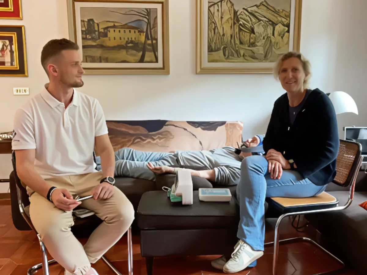
Cells are collected from the patient's own bone marrow (autologous, approximately 3-5 ml under local anaesthesia) or sourced from a certified donor (allogeneic), depending on individual clinical indications. Cells are expanded, quality-controlled and tested in our certified Warsaw laboratory. Administration is performed by percutaneous injection directly to the fracture site under imaging guidance.
Biological Mechanisms
How This Helps in a Non-Union Fracture
In non-union fractures, the osteogenic response has failed. The fracture site lacks sufficient progenitor cells, growth factor signalling and vascular supply to form new bone. MSCs directly address all three deficits: they provide the cellular substrate for bone formation, secrete the molecular signals needed to drive osteogenesis, and stimulate new blood vessel growth to sustain the healing tissue.
Your Medical Board
The exact combination, dosage, sequencing and delivery method of all five components is determined individually by our medical board for each patient. No two treatment protocols are identical. Your programme is constructed based on your specific fracture type, location, vascularity, metabolic profile, prior surgical history and clinical priorities.
Your protocol is designed individually. Speak with our medical team to understand what your personalised program would include.
Request ConsultationPatient Journey
Your case is reviewed remotely by our physician team. We assess your imaging, fracture history, prior surgical interventions and treatment goals. This consultation is free and carries no obligation.
A detailed review of all medical documentation, including current imaging (X-ray, CT), infection markers, vascular assessment and metabolic panel. Our medical board evaluates eligibility, confirms safety parameters and designs your personalised therapeutic protocol.
Your cells are collected, isolated, expanded and quality-tested in our certified Warsaw laboratory. Each batch receives a full traceability certificate. This stage typically takes 2–3 weeks.
Cellular material is delivered by percutaneous injection directly to the fracture site under imaging guidance — no open surgery, no general anaesthesia. Treatment is administered at our Warsaw clinic. Airport transfers, accommodation and visa support are included in the programme.
Structured rehabilitation programme with our specialist, adapted to your fracture location and current weight-bearing status. Progressive loading protocols are calibrated to support biological healing while preventing re-injury.
Your dedicated coordinator monitors radiographic progress, functional recovery and overall healing trajectory. Serial imaging is reviewed at defined intervals. A medical-grade wearable bracelet supports continuous health tracking regardless of your location.
The first step is free. Request a medical consultation and our medical consultant will contact you within 24 hours.
Request ConsultationSafety Profile
Cellular therapy for bone healing is considered safe when delivered under proper medical supervision and according to validated protocols. In our practice, the procedure is well-tolerated by the majority of patients.
Temporary mild reactions — such as local discomfort at the injection site, slight swelling or low-grade temperature — may occur in a minority of patients. These are typically short-lived and resolve within 48–72 hours.
A final medical assessment is performed on-site before every treatment session. If a patient's status has changed — including evidence of new infection or metabolic decompensation — the programme may be temporarily modified or postponed for safety reasons.
All contraindications are evaluated individually. A contraindication in one clinical context does not necessarily preclude treatment in a different context — this is always determined by physician assessment.
Standard Contraindications
Active infection at the fracture site (requires staged approach — infection control first)
Active malignancy or ongoing chemotherapy / radiotherapy
Severe uncontrolled diabetes (HbA1c > 10%) — requires metabolic optimisation before treatment
Pregnancy
Active osteomyelitis with systemic sepsis
Post-Treatment
Dedicated rehabilitation specialist
monitors weight-bearing progression, range of motion and functional recovery
Progressive loading protocol
carefully calibrated to support bone consolidation without overloading the healing fracture
Medical-grade wearable monitoring
continuous physiological data collection supporting clinical decision-making
Serial imaging review
radiographic assessment at defined intervals to confirm healing progression and callus maturation
Long-term coordinator support
proactive check-ins, clinical guidance and response to any changes in healing status
Bone consolidation is a biological process that continues for months after cellular delivery. The post-treatment period requires structured monitoring, progressive mechanical loading and metabolic optimisation. Imaging milestones guide all decisions about weight-bearing progression and return to full activity.
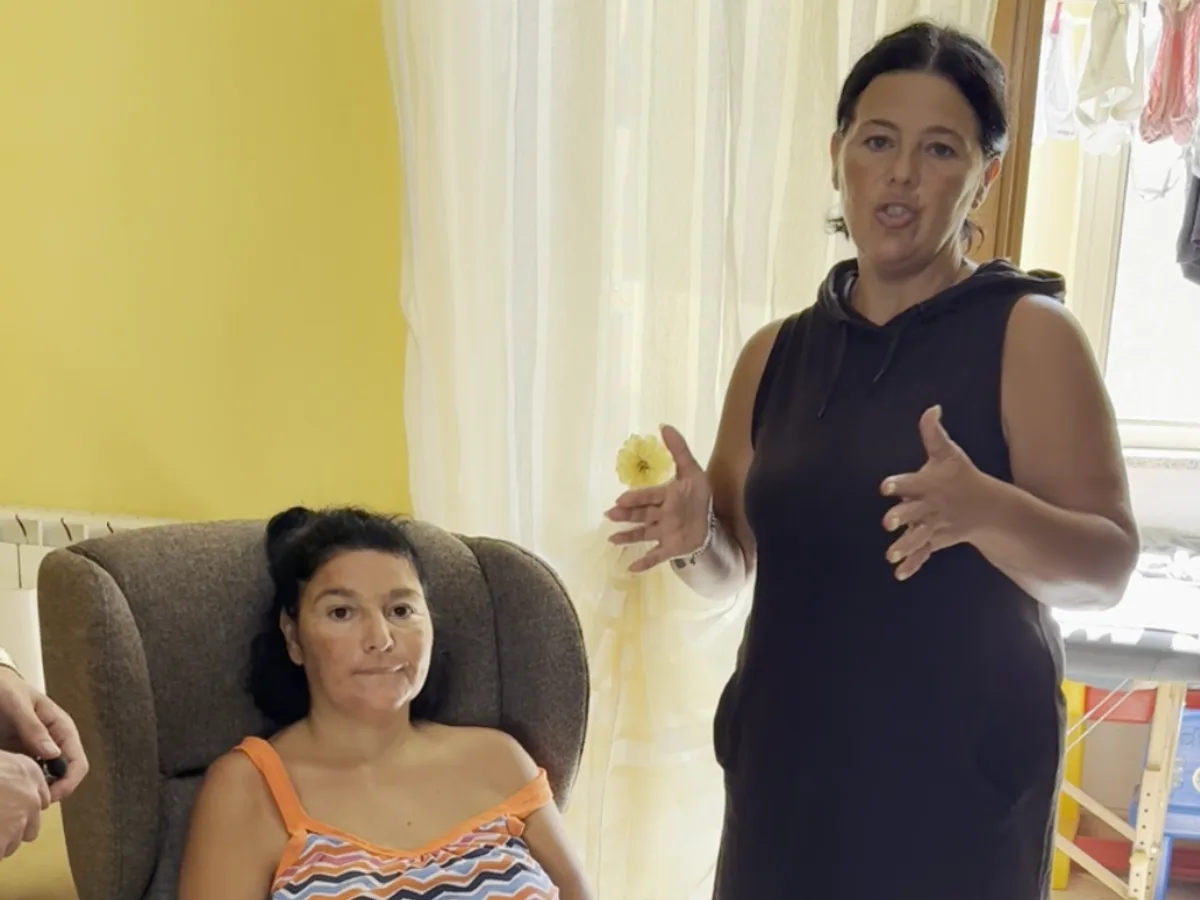
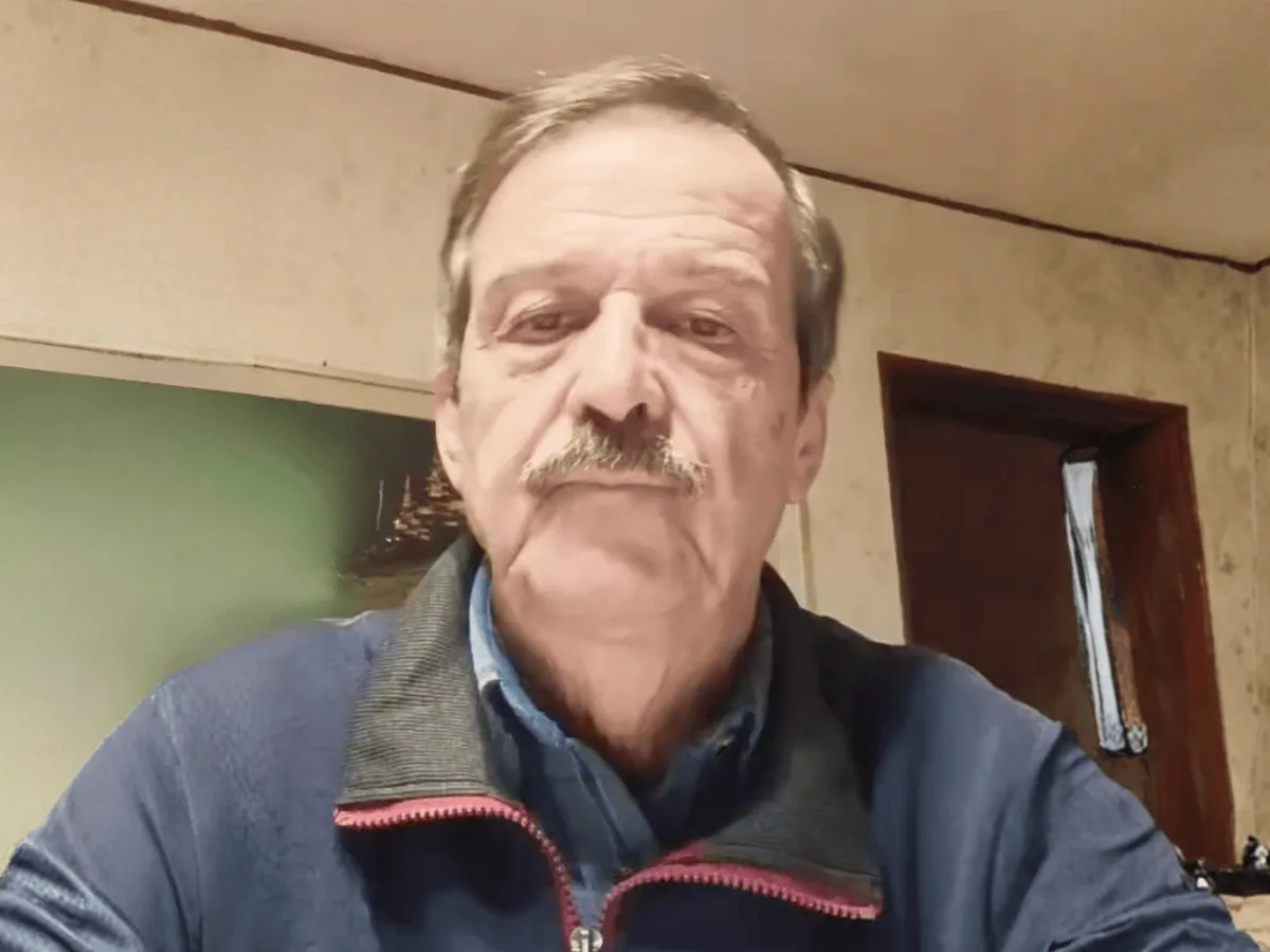
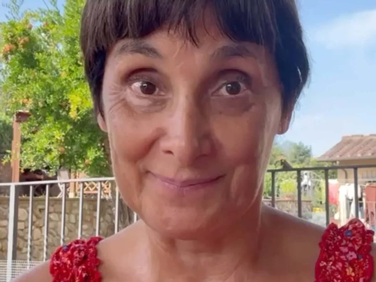
Patient Stories
“Eighteen months, two surgeries, and my tibia still had not healed. They were planning a third operation with a bone graft. I went to Warsaw instead. Within a few months the imaging showed new bone forming across the gap. I am walking without crutches for the first time in over two years.”
Patient
Atrophic tibial non-union · Germany
Every case is assessed individually by our physician team. Request a consultation to discuss your specific situation with our physician team.
Request ConsultationPatient Cases
Documented treatment outcomes recorded by the BioCells Medical team after personalised regenerative medicine protocols.
Get Started
If you or someone you care for has a fracture that has failed to heal — despite surgery, bone grafting or prolonged immobilisation — our medical team is available for a free, no-obligation medical consultation based on your imaging, treatment history and clinical profile.
We review every inquiry personally. You will speak with a physician, not an administrator.
Submit your case online or by phone
Our medical consultant contacts you to review your documents
The medical board presents your personalised treatment plan
Request a Consultation
Tell us about your condition. Our medical consultant will contact you within 24 hours to review your documents.
Open Consultation FormMultilingual coordination — English, Italian, French, Russian, Polish
Evidence Base
Our clinical approach is informed by and consistent with published research in the field of regenerative medicine.